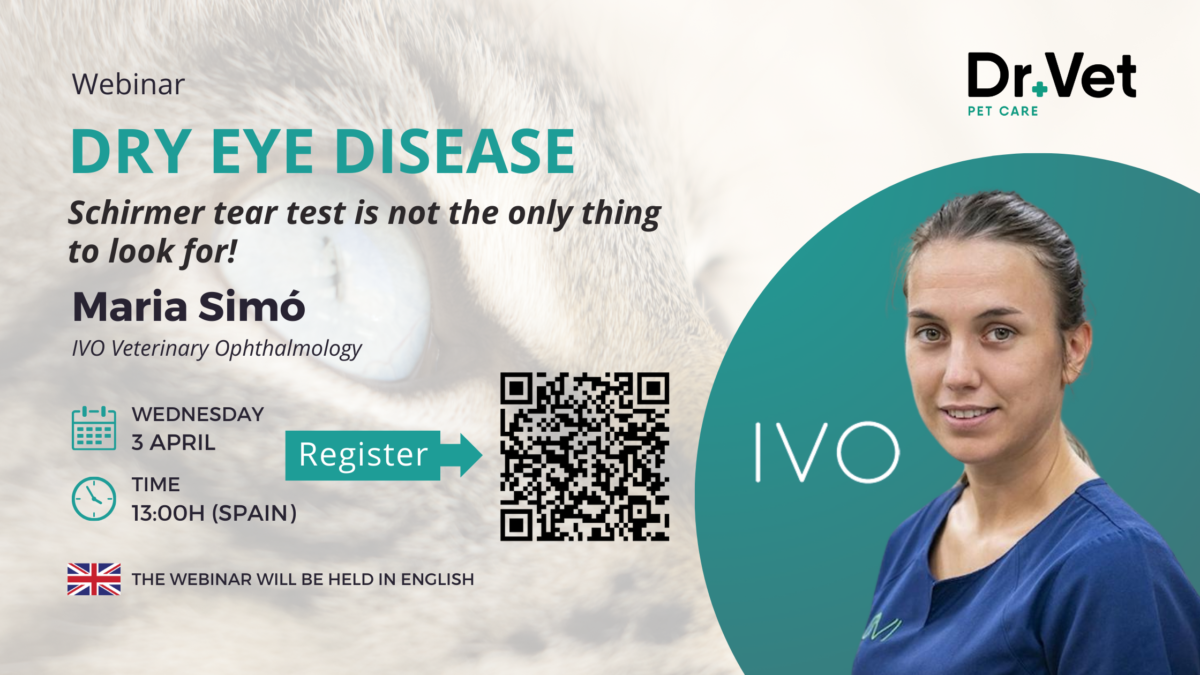
El pasado miércoles 3 de abril, Dr+Vet tuvimos el honor de recibir a la destacada oftalmóloga veterinaria María Simó en nuestro primer webinar sobre Queratoconjuntivitis Seca (KCS) en perros. Durante este evento virtual, María Simó compartió su conocimiento y experiencia en el diagnóstico y tratamiento de esta enfermedad ocular común pero a menudo subestimada. Hoy, por fin podemos anunciar que el video completo, con subtítulos en inglés (y otros idiomas de forma automática), está disponible para su visualización en nuestro blog y nuestro canal de YouTube! Adjunto a este artículo, encontrarás el enlace directo al video para que puedas acceder a él y profundizar en los conocimientos compartidos por María Simó.
Sobre María Simó:
María Simó es una reconocida oftalmóloga veterinaria con una sólida formación y una amplia experiencia en el campo de la oftalmología veterinaria. Graduada en Medicina Veterinaria por la Universidad Autónoma de Barcelona, María ha completado diversos cursos de postgrado y se desempeña como oftalmóloga en el prestigioso Instituto Veterinario Oftalmológico (IVO) de Barcelona.
Contenido del Webinar:
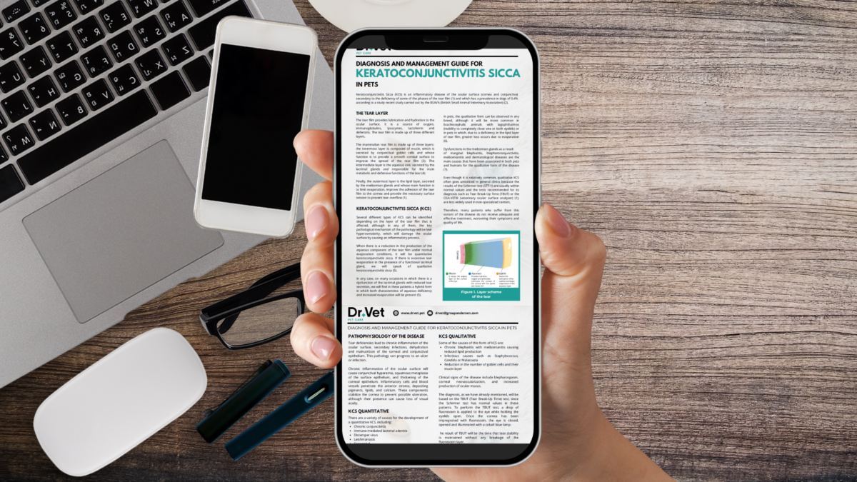
María Simó nos habló de los fundamentos de la Queratoconjuntivitis Seca, desde los métodos de diagnóstico hasta las opciones de tratamiento más efectivas, explorando en detalle cómo abordar esta enfermedad de manera integral.
Una de las principales conclusiones del webinar fue la importancia de no depender únicamente del Test de Schirmer para diagnosticar KCS. María Simó enfatizó la necesidad de una evaluación completa y detallada, así como el uso de herramientas diagnósticas adicionales para un correcto diagnóstico, ya que, sin toda la información se pueden recetar tratamientos ineficaces. También remarcó la importancia de derivar casos que no terminan de resolver correctamente para poder evaluarlos al completo.
Agradecemos a todos los participantes su apoyo
El webinar sobre Queratoconjuntivitis Seca con María Simó fue un gran éxito, y queremos agradecer a todos los que se unieron a nosotros en este evento educativo. Esperamos que este recurso continúe siendo una fuente valiosa de información para todos los veterinarios interesados en la oftalmología veterinaria. No dudes en consultar el video completo y compartirlo con tus colegas.
¡Sigue atento a nuestras redes sociales y sitio web para obtener más información sobre futuros eventos y recursos educativos de Dr+Vet!